Ventricular Tachycardia
Ventricular tachycardia (VT) is a potentially fatal cardiac arrhythmia that can arise from aberrant electrical conduction in the heart. It is usually due to scar tissue from a previous myocardial infarction (heart attack). VT can cause sudden cardiac arrest and is therefore life threatening.
VT is treated through medical management (drugs), catheter ablation (radiofrequency ablation), and with an implantable cardiac defibrillator (ICD). However, the recurrence rate with these therapies can reach 50% at one year.
Noninvasive Cardiac Radioablation
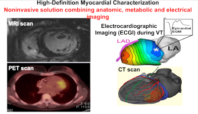
Noninvasive cardiac radioablation combines noninvasive imaging (CT, MRI, PET, and ECGI) to identify and target a VT arrhythmia with stereotactic radiosurgery. The Wash U program was developed and is lead by our collaborators Dr. Phill Cuculich, an electrophysiologist, and Dr. Cliff Robinson, a radiation oncologist.
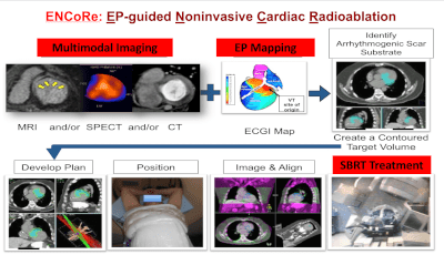
The Wash U team has published a series of clinical studies showing the promise of this technology in patients. CORAL is collaborating to help improve the efficiency and accuracy of noninvasive cardiac radioablation. Specifically, we are interested in software and algorithms to improve target definition, improve delivery accuracy in the presence of motion, and to help disseminate and test this technology more widely.